Ankle-Brachial Index, CT Angiography, and Proximal Tibial Traction for Gunshot Femoral Fracture
Main Text
Table of Contents
This video demonstrates an algorithm for evaluating suspected vascular injury secondary to penetrating extremity trauma. Descriptions of how to perform an arterial-brachial index (ABI) and arterial-pulse index (API) are reviewed, along with criteria to determine if a computed tomography angiography (CTA) is indicated. Relevant imaging is reviewed with a radiology resident with descriptions of how to systematically assess the scans for injury. The technique for a tibial traction pin, a temporizing measure for long bone fractures, is described.
Penetrating extremity trauma (from ballistic injuries and stab wounds) is a common injury pattern seen in urban trauma centers. It is imperative for medical professionals to understand the algorithm for evaluating vascular injury in these patients.
The patient discussed in this case is a 42-year-old man with no significant past medical history who sustained a gunshot wound to his left lower extremity. He was transferred by emergency medical services from the field to the emergency department where he was evaluated by the trauma surgery service. His primary survey was intact upon arrival. After life-threatening injuries were ruled out with the primary exam, a more detailed “secondary” physical exam was conducted.
A thorough physical exam demonstrated two lacerations, presumed to be from the ballistic injury to the medial and lateral left thigh. There was an obvious deformity, swelling, and associated pain of the proximal left lower extremity. The compartments of the patient’s thigh were appropriately swollen but soft. Pulses of the dorsalis pedis and posterior tibial arteries were palpable.
Plain film imaging of the left lower extremity demonstrated a spiral fracture of the left femur. There was a drill-type fracture of the distal femur. A subsequent computed tomography angiography (CTA) re-demonstrated the spiral femur fracture and noted adequate distal vascular runoff.
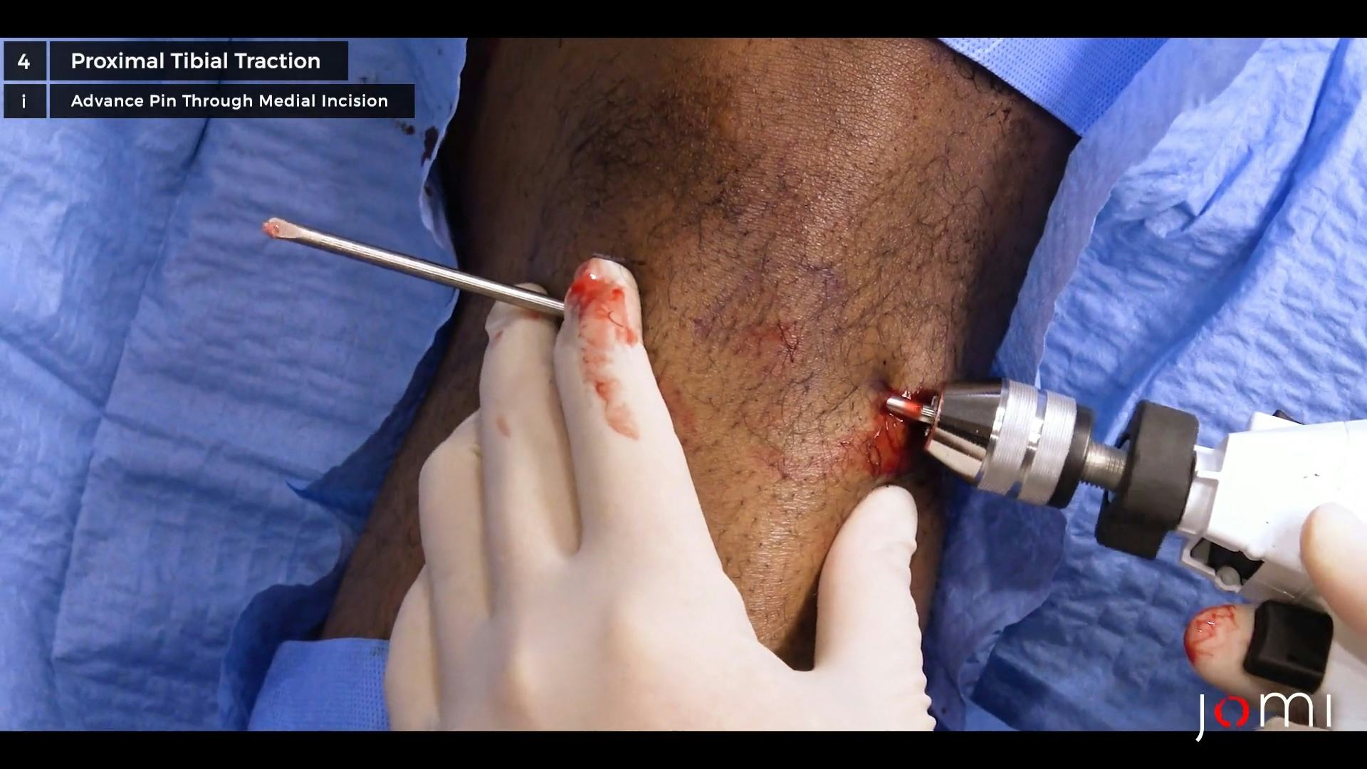
Penetrating trauma, such as a ballistic injury, poses threat to osseous and neurovascular structures. Upon arrival, patients are ultimately triaged by means of physical examination and non-invasive modalities. All patients who arrive with a penetrating injury have their vaccination status assessed and are provided with the tetanus vaccine if they are not up to date on their vaccination. In this particular patient, lower extremity vascular status was initially assessed upon secondary survey with palpation of the dorsalis pedis and posterior tibial pulses. An arterial-brachial index (ABI) and arterial-pulse index (API) were utilized to further assess the adequacy of limb perfusion. The API, which evaluates the relative perfusion of the injured and non-injured extremity, was obtained and noted to be 0.87. A CTA was subsequently completed given the API less than 0.9. Re-demonstration of the spiral femur fracture was noted on CT. There was no evidence of contrast extravasation, pooling, vasospasm, or pseudoaneurysm on imaging suggesting adequate distal runoff. After ruling out vascular injury, the orthopaedic surgery team was consulted for further care. Diaphyseal femur fractures are prone to shortening of the distal fracture fragment due to spasm of the surrounding musculature. To temporize the fracture and offer pain control, a proximal tibial traction pin was applied.
The orthopaedic surgery team temporized the femur fracture with skeletal traction. This traction may be performed either in the distal femur or proximal tibia. Given the distal extension of the femur fracture and after confirming a ligamentous stable knee, proximal tibial traction was applied. This procedure was again completed for analgesia. The patient was later taken to the operating room for intramedullary fixation following adequate resuscitation.
This patient presented with a single ballistic injury to his left lower extremity. The incidence of vascular injury secondary to penetrating trauma has been variably reported in the literature. For the lower extremities, the incidence ranges from 3–25%, and injury most commonly occurs to the femoral or popliteal vessels.1–3 While his peripheral pulses were palpable, penetrating injuries should be further evaluated by validated, non-invasive measures. His API was less than 0.9, and therefore, a CTA was completed per our institutional and consensus algorithm. Fortunately, there was no evidence of vascular injury on further imaging. The orthopedic surgery service was consulted and a proximal tibial traction pin was placed prior to operative intervention.
The ABI and API are validated, non-invasive means for evaluating subtle vascular injuries that do not have any “hard signs” of vascular injury. These hard signs include pulseless extremity, expanding hematoma, pulsatile bleeding, or signs of a pseudoaneurysm including a palpable thrill and audible bruit. Any patient that has one of these “hard signs” in the setting of any kind of trauma should be taken to the operating room for urgent exploration.1, 4
In the absence of a hard sign of vascular injury but with a penetrating injury mechanism concerning vascular pathology, an ABI or API should be ascertained. As shown in the video, an ABI is evaluated by placing the sphygmomanometer on a patient’s calf and assessing the systolic blood pressure at the level of the dorsalis pedis or posterior tibial artery. This is then compared to the systolic blood pressure from the ipsilateral brachial artery. Two commonly used, though non-validated, ways to quickly estimate an ABI are to use the systolic blood pressure from the automated blood pressure cuff or to use the radial artery (as demonstrated in the video) as a proxy for the brachial artery pressure. While these can help provide an idea of what an ABI would be, they are not adequate replacements for a true ABI. If the patient does not have an ipsilateral extremity from a devastating blast injury or prior amputation, an alternative to the ABI is the API in which the injured limb is compared to the contralateral extremity. The API is measured as a ratio of the pressure at which the pulse (e.g. in our patient the posterior tibial) returns via Doppler relative to that of the contralateral limb.
An ABI less than 0.9 has traditionally been used as a threshold for if a patient should require further testing for vascular injury. At this level, the ABI has a 95% sensitivity and 97% specificity for assessing the clinically-significant vascular injury.1 Guidelines from the two major associations of trauma surgeons (EAST and WEST) agree that patients with an ABI greater than 0.9 can be safely discharged home if no other concern for injury is present as it has been shown that only 5.5% of these patients will return with complications nearly all of which will be wound complications.1, 4, 5 More recent work suggests that for penetrating trauma to the extremities this may still be too high of a threshold and that clinically significant vascular injury is not seen with an ABI >0.7.6
Historically, all patients with concern for vascular injury would require on table angiography to evaluate the vessels of the lower extremity.1 This is a procedure by which a catheter would be placed in the femoral artery of the contralateral lower extremity advanced proximally to the aortic bifurcation and contrast would be evaluated as it was directed down into the vessels of the lower extremity. With the advent of CTA, this invasive procedure has been replaced with non-invasive imaging while maintaining 100% specificity and sensitivity for vascular injury and significantly reducing cost for both patients and the hospital.1
Interestingly, when reviewing the mechanism of the spiral fracture seen on the patient’s X-ray and CT, the bullet was not likely the source of the fracture. Ballistic fracture patterns are more commonly comminuted or drill type. Upon further history, the patient noted that he fell after being struck by the bullet. A twisting mechanism, while falling, could explain this spiral fracture pattern. Given the lack of retained projectile and two wounds on the medial and lateral aspect of his thigh, this patient sustained a through-and-through ballistic injury.
In this case, the patient proceeded to the operating room the next morning where he underwent open reduction with internal fixation. He was evaluated by physical therapy the following day and was able to be discharged home with instructions to follow up as an outpatient and work with outpatient physical therapy on the second day of his hospital stay.
No special equipment was used during this case.
Nothing to disclose.
The patient referred to in this video article has given their informed consent to be filmed and is aware that information and images will be published online.
The authors received no funding for this work.
Citations
- Fox N, Rajani RR, Bokhari F, et al. Evaluation and management of penetrating lower extremity arterial trauma: an Eastern Association for the Surgery of Trauma practice management guideline. J Trauma Acute Care Surg. 2012;73:S315-S320. doi:10.1097/TA.0b013e31827018e4.
- deSouza IS, Benabbas R, McKee S, et al. Accuracy of physical examination, ankle-brachial index, and ultrasonography in the diagnosis of arterial injury in patients with penetrating extremity trauma: a systematic review and meta-analysis. Acad Emerg Med. 2017 Aug;24(8):994-1017. doi:10.1111/acem.13227.
- Weinberg DS, Scarcella NR, Napora JK, Vallier HA. Can vascular injury be appropriately assessed with physical examination after knee dislocation? Clin Orthop Relat Res. 2016 Jun;474(6):1453-8. doi:10.1007/s11999-016-4730-6.
- Feliciano DV, Moore FA, Moore EE, et al. Evaluation and management of peripheral vascular injury. Part 1. Western Trauma Association/critical decisions in trauma. J Trauma. 2011 Jun;70(6):1551-6. doi:10.1097/TA.0b013e31821b5bdd.
- Sadjadi J, Cureton EL, Dozier KC, Kwan RO, Victorino GP. Expedited treatment of lower extremity gunshot wounds. J Am Coll Surg. 2009 Dec;209(6):740-5. doi:10.1016/j.jamcollsurg.2009.09.010.
- Hemingway J, Adjei E, Desikan S, et al. Re-evaluating the safety and effectiveness of the 0.9 ankle-brachial index threshold in penetrating lower extremity trauma. J Vasc Surg. 2020;72(4):1305-1311.e1. doi:10.1016/j.jvs.2020.01.051.
Cite this article
Kent JR, Jeffries J, Straszewski A, Wilson KL. Ankle-brachial index, CT angiography, and proximal tibial traction for gunshot femoral fracture. J Med Insight. 2023;2023(299.7). doi:10.24296/jomi/299.7.